“Are you still teaching the three-sided dressing?”
This question was recently asked to one of our instructors during a training session for first responders in isolated regions. It highlighted the ongoing confusion due to entrenched reflexes and assumptions, despite evolving recommendations. Today, multiple guidelines, based on evidence from recognized organizations, suggest different approaches.
This prompted a thorough review of current literature and guidelines to clarify best practices.
Let's start with a realistic scenario
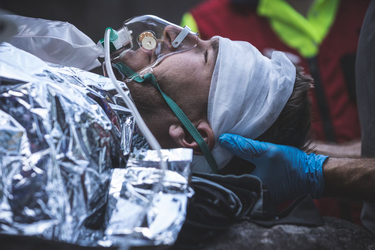
 A mountain biker falls on a remote trail, sustaining a severe injury by landing on a sharply cut spruce stump. Instinctively pulling the branch out upon rising, he leaves a gaping chest wound.
With each inhalation, a whistling sound is audible. Quickly, he becomes dyspneic, restless, and confused. A teammate, equipped with a first aid kit and trained in advanced wilderness first aid, prepares a dressing with gauze and a survival blanket but remains uncertain: “Should I seal three sides or four?”
RECOGNIZING an Open Pneumothorax
An open chest wound can quickly progress to a tension pneumothorax or hemothorax. Look for these signs:
- Penetrating chest injury, often with air leakage
- Sucking or whistling sounds during breathing
- Respiratory distress or paradoxical chest movements
- Cyanosis of lips or extremities
- Diminished or absent breath sounds on the injured side
- Altered mental status, agitation, or signs of shock
ACTION
DO NOT REMOVE AN OBJECT IMPALED IN THE CHEST
. This could worsen bleeding or pulmonary damage.
The Three-Sided Dressing: Still Relevant or Outdated?
For decades, applying an occlusive dressing taped on three sides has been taught as a standard treatment for open pneumothorax, creating an improvised one-way valve allowing air to escape during expiration while blocking air entry during inhalation.
Currently, this method is debated in various professional circles. Although still recommended by some sources (e.g., ATLS 10th edition), many specialized organizations have abandoned it in favor of more reliable and safer field methods. Direct scientific evidence against this technique remains limited, often derived from animal models or indirect clinical observations.
Main identified limitations:
- Variable effectiveness: sweat, blood, hair, and movement can impair the improvised valve's function.
- Risk of blockage: plastic can seal tightly, preventing air escape.
- Potential deterioration: improper monitoring may lead to tension pneumothorax.
|
ORGANIZATION |
CURRENT RECOMMENDATION |
|
TCCC (COMBAT CARE) |
Commercial chest seal with valve preferred; if unavailable, fully sealed with active monitoring. Perform burping if deterioration occurs. |
|
ATLS (10th ED.) |
Three-sided dressing still recommended without chest drainage. |
|
WMS (WILDERNESS MEDICAL SOCIETY) |
Fully sealed or valve-integrated dressings recommended; three-sided no longer advised. |
|
ITLS / NAEMT / PHTLS |
Three-sided dressing removed; recommend commercial valve seal or four-sided dressing with close monitoring. |
| ERC (EUROPE)) |
If no valve available, sometimes leave the wound open. |
|
RED CROSS / ST. JOHN AMBULANCE |
Recent guides favor fully sealed dressing with active clinical monitoring; caution against improvised dressings. |
🔷 SIRIUSMEDx Position
At SIRIUSMEDx, we primarily recommend:
- Commercial chest seals with integrated one-way valves when available, as these provide superior effectiveness in real conditions (sweat, hair, movement).
- If valves are unavailable, a fully occlusive dressing sealed on four sides is advised with continuous clinical monitoring.
We recognize some training contexts may still teach the three-sided dressing, but safe application requires technical precision and careful clinical observation.
Our emphasis is less on the number of taped sides and more on the responder’s ability to observe, assess, and respond.
- Think clinically rather than technically
- Understand your actions
- Monitor respiratory impacts
- Master critical simple maneuvers (like burping, explained below)
- Initiate rapid and well-documented evacuation
Application of Occlusive Dressing – 3-sided vs. 4-sided
Four-Sided Dressing (Fully Occlusive)
Objective: Completely seal the wound to prevent air entry while maintaining strict monitoring.
Materials:
- Sterile gauze
- Plastic film (ziplock bag, plastic wrap, Mylar)
- Medical adhesive tape (duct tape, elastoplast)
Steps:
- Clean surrounding skin (blood, sweat, dirt).
- Apply slightly damp gauze over the wound.
- Cover with plastic film without adhering directly to the gauze.
- Seal all four edges.
- Avoid direct plastic contact with the wound; leave an air pocket or fold.
- Watch closely for signs of tension pneumothorax. If suspected → burp or decompress if qualified.
Three-Sided Dressing (Improvised Valve)
Objective: Allow passive air escape on expiration while blocking air entry on inspiration.
Steps:
- Prepare the skin as above.
- Place gauze followed by plastic film.
- Tape three sides only, leaving one lower corner open.
- Orient open corner downward.
- Create a small air tunnel centrally to avoid suction.
- Frequently reassess; burp if deterioration occurs.
Caution: This method is technically demanding and can also cause tension pneumothorax.
Monitoring Closely After Applying Occlusive Dressing:
Recognizing Tension Pneumothorax
An occluded chest wound may progress to tension pneumothorax regardless of dressing type—air accumulation compresses the affected lung, shifting mediastinal structures and threatening breathing and circulation.
|
CLINICAL SIGNS |
WHAT TO LOOK FOR ? |
|
Breathing quality |
Is the breathing asymmetrical, shallow, or ineffective on one side? This could indicate that one lung is being compressed by a developing tension pneumothorax. |
|
Skin and lip colour |
skin turning from pink to grey or bluish (cyanosis) is a sign of poor oxygenation. |
|
Mental status |
agitation, confusion, or drowsiness may indicate cerebral hypoxia. |
|
Pulse |
a rapid, thready pulse may be an early indicator of clinical deterioration, compensated shock, or mediastinal compression. |
|
Chest symmetry: |
Absent breath sounds or reduced chest expansion on one side may suggest air accumulation under pressure. |
|
Tracheal deviation
|
Tracheal deviation: a late and uncommon sign (present in only 2.3% of cases) suggesting a shift of the mediastinum away from the affected side, typically seen in advanced tension pneumothorax. |
Burping: A Simple Action, but a Decision Based on Rigorous Clinical Assessment
Burping (temporarily lifting a dressing corner) relies on specific clinical signs, not intuition.
Consider burping if:
- Sudden increase in respiratory distress
- Increased chest asymmetry
- Altered consciousness
- Cyanosis or unexplained hypotension
- Palpable tension or hyperresonance (if qualified)
Safe Burping Technique:
- Gently lift one dressing corner for 1-2 seconds.
- Listen for escaping air.
- Immediately reseal after air release.
- Resume close monitoring.
This action can save time—and lives—by relieving pressure and improving ventilation while awaiting needle decompression (if qualified) or rapid medical evacuation.”
Needle Decompression: A Reserved Medical Act, but a Shared Decision Reserved for trained professionals
Even though this procedure is reserved for trained and authorized personnel, the decision to consider it should first be based on an objective clinical assessment. A responder must be able to:
- Recognize the clear clinical criteria of a tension pneumothorax
- Anticipate clinical progression in the absence of a decompression tool
- Communicate clear clinical data to remote medical support teams
Needle Decompression – For Qualified Personnel:
🔸 Conditions:
- Legal scope (nurse, paramedic, medic, physician)
- Validated training
- Under medical protocol or supervision
🔸 Site:
- 5th intercostal space, anterior axillary line
- 2nd intercostal space, mid-clavicular line
🔸 Material:
- 14G catheter, ≥8 cm length
- Commercial decompression needle (ARS, PneumoDart, etc.)
This is a life-saving intervention—not a reflex. It should only be performed with strong clinical justification.
 vacuation of a Patient with a Penetrating Chest Wound: A Top Priority
Even when clinically stabilized, a patient with a penetrating chest wound must be evacuated without delay.
🔻 evolving risks:
- Tension pneumothorax
- Hemothorax
- Pulmonary contusion
- Cardiac tamponade
✅ While awaiting evacuation:
- Semi-seated position if tolerated
- If not possible, lateral decubitus on the injured side
- Oxygen if available
- Frequent reassessments
Special Considerations for Gunshot Chest Wounds
Projectile Chest Injuries (e.g., firearms, shrapnel) Require Special Attention:
- Always assess for both an entry and an exit wound. A projectile can completely traverse the thoracic cavity. Each wound may serve as an entry point for air or a pathway for air escape. Both should be occluded as soon as possible.
- If two occlusive dressings are available, use one for each wound. If only one is available, prioritize the wound with active air leak (audible airflow, bloody froth, whistling on inhalation).
Air Transport: Be Alert to Pressure Changes!
At altitude, as atmospheric pressure drops, intrapleural air (the space between the collapsed lung and chest wall) expands, increasing the risk of clinical decompensation.
🎯 Precautions :
- Inform the transport team: include details about the chest seal and clinical evolution.
- Pre-flight burping if signs of tension are present
- Keep emergency intervention equipment ready
- Adjust flight altitude if necessary
Clinical Summary for Handover (SIRIUSMEDx Template)
- Type of injury: open chest wound, right flank
- Time of incident: [HH:MM]
- Interventions: sealed dressing, burping ×1
- Clinical signs: RR, HR, SpO₂, level of consciousness
- Current status: stable / in distress / decompensated
- Needs: urgent evacuation, medical oversight
Responder Scope of Practice – Summary Table
|
Level |
Permitted Actions |
Primary Role |
|
First Aider (WFA, AWFA, WFR) |
Dressing application, burping |
Stabilize, monitor, initiate evacuation |
|
Advanced (Paramedic, Nurse) |
Dressing, burping, decompression if qualified |
Actively intervene and document |
|
Physician |
Dressing, burping, decompression, chest drain |
Assess, coordinate, anticipate |
Examples of Useful Commercial Chest Seals
|
Model |
Advantages |
Suitable Settings |
|
SAM® Chest Seal |
Valve, highly adhesive |
Military, industrial, hunting |
|
HALO® |
Adheres with sweat/blood |
Military, industrial, hunting |
|
Russell® |
Visual valve |
Pediatric, tactical |
|
Improvised (Mylar + tape) |
Accessible and adaptable |
Isolated or austere regions |
Conclusion: Choosing the Best Approach for the Context
The debate over three-sided, four-sided, or valve chest seals reflects the evolution of clinical practices and the importance of staying current. There is rarely a one-size-fits-all answer, but better decisions can be made depending on the context, available materials, and the responder's level of competence.
Teaching the four-sided sealed dressing has the advantage of simplifying the approach, reducing technical errors, and aligning with expert recommendations. However, beyond technique, what truly matters is understanding what you're doing, why you're doing it, and how to monitor the effects.
Regardless of the dressing used, clinical monitoring remains the cornerstone: recognizing tension pneumothorax, intervening appropriately (burping, decompression), and initiating a rapid, well-prepared evacuation.
At SIRIUSMEDx, we favour methods proven effective in the field—but more importantly, we train responders to assess, adapt, and justify their decisions. Because in remote environments, the best decisions are often those made with insight, not reflex.